Every year, thousands of Israelis suffer from kidney stones that lead to excruciating pain. These stones are small, but they can cause significant issues. Some of them are spontaneously expelled from the body, while others require various methods of removal.
More stories:
New techniques allow for precise treatment of the issue with a quick recovery period, despite involving surgical procedures.
Kidney stones form when urine becomes too concentrated due to the presence of high amounts of salt content. When urine is concentrated, crystals accumulate in it, most of which are made of salts, forming what are referred to as stones.
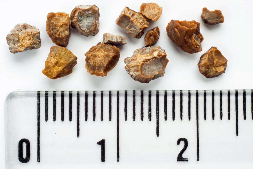
The kidney stones are crystalline deposits of minerals that dissolve in urine. The stone mostly forms in the kidney or the ureter, which is the tube that carries urine from the kidneys to the bladder. Stones can vary in size; some are as small as a grain of sand, while larger ones can be the size of a cherry.
If located in the kidneys, they’re called "nephrolithiasis," and if they’re found in other parts of the urinary tract, they’re called "ureterolithiasis." Most kidney stones are extremely small; the largest one found to date weighed 1.36 kg.
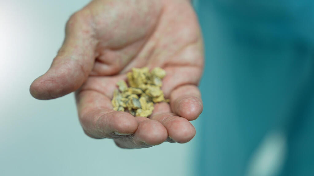
They can come in various colors, but most are yellow-brown in hue. The majority of these stones are formed as a single unit and are also passed as such. So far, about 200 components have been identified in different types of stones, with the most common being calcium oxalate, phosphorus, uric acid and cystine.
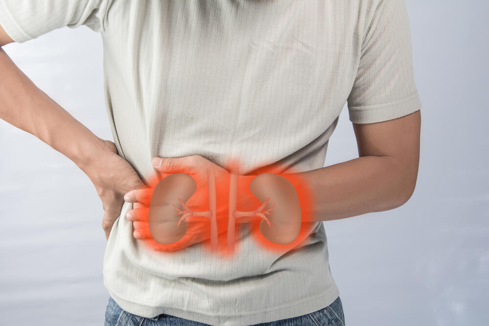
In most cases, kidney stones pass in the urine without causing noticeable symptoms, but if a relatively large stone – 2 mm or more in size – forms, it can cause a blockage in the ureter. This results in the tube’s dilation, obstruction of urine flow, and severe, acute pain typically felt in the lower back, lower abdomen, and groin.
These severe abdominal pains are medically referred to as colic and can also lead to nausea and vomiting. The stone can also occasionally injure the ureter, causing blood to appear in the urine.
Diagnosis of kidney stones usually begins with the patient's complaints of pain and hematuria (presence of blood in urine) during a urinalysis. An ultrasound examination can show kidney swelling (hydronephrosis) caused by the stone blocking the urinary tract or reveal a silhouette indicating the presence of a kidney stone. If the stone contains a sufficient amount of calcium, it can also be seen in an X-ray.
If the stone is small and causes severe pain, the patient may be hospitalized and receive IV hydration therapy in order to increase the amount of urine produced and to help pass the stones naturally. Alongside that, the patient may be administered pain-relieving medications.
If the stone remains in place, non-invasive treatment can be attempted using shockwave lithotripsy. "The treatment involves a specialized device that generates focused sound waves targeted at the stone under the guidance of X-ray," explains Professor David Lifshitz, head of the Rabin Medical Center’s Minimally Invasive Urology unit.
"The goal is to break the stone into small fragments that can be passed on their own. The advantage of the procedure is that it’s non-invasive. The downside is that not all stones respond to the energy of the sound waves."
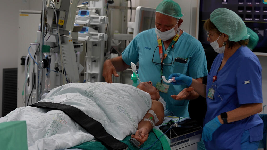
Other cases require surgical treatment under anesthesia. The two relevant options are ureteroscopy and percutaneous nephrolithotomy. "In ureteroscopy, we insert a telescope into the ureter through the urethra," says Prof. Lifshitz. "Looking through the telescope, and using a laser, we break the stone into fragments and remove the pieces.”
“A more invasive approach is percutaneous nephrolithotomy,” he explains. “In this approach, designed for larger kidney stones, an artificial path is created to reach the kidney – a small hole in the hip through which a telescope is inserted, after which the stone is ground and extracted using a special device."
After the procedure, a stent remains – a thin silicone tube that passes between the ureter and the kidney. "Patients may complain about irritation and discomfort," says Lifshitz, "but the recovery is quick. After a ureteroscopy, the patient is released home on the same day and can resume regular activities.”
“After a percutaneous nephrolithotomy surgery, it’s recommended to avoid exertion for two weeks, but the patient is discharged the day after the surgery and can move around. They aren’t bound to stay in bed,” he added.
Every patient suffering from kidney stones undergoes diagnoses aimed at identifying the cause of their formation. "It's a chronic disease with a 50% recurrence rate," says Lifshitz. "Therefore, it's important to conduct metabolic testing for patients, including analyzing the composition of the extracted stone and collecting urine samples over 24 hours for salt analysis.”
“By doing this, we can piece together and understand the underlying causes, and design a preventive treatment, dietary recommendations and pharmacological intervention that could prevent stone recurrence," he added.


