


A chest CT scan is usually ordered to answer one medical question, but the scanner does not distinguish between the organ that prompted the referral and everything around it. Along with the lungs, the scan also captures the heart, bones, part of the liver and the esophagus, each of which may contain information no one was looking for.
A new study published in Nature Medicine showed that an artificial intelligence system can extract signs of esophageal cancer from that information, even in scans performed without contrast material and not intended to look for a tumor in the esophagus.
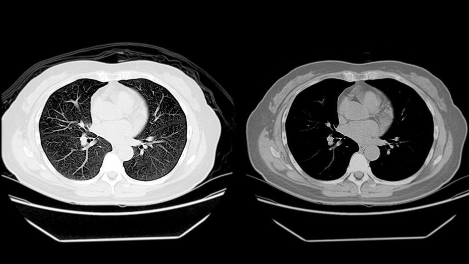
If the approach proves itself in further studies, the implications could be broad: Instead of ordering a new test every time another need arises, medicine could make smarter use of the millions of scans already being performed.
Radiology has a name for this phenomenon. “We call them incidental or ancillary findings, or, if we are talking about doing one test and wanting to use it to identify another disease, ‘opportunistic screening,’” explains Dr. Arnon Makori, head of imaging at Assuta Medical Centers and a diagnostic radiologist specializing in abdominal imaging.
One example is low-dose CT, which is used to screen people at risk for lung cancer. “We look at the lungs to detect lung cancer, but we also have an opportunity here to identify additional disease, associated disease or incidental disease,” Makori says. According to him, the same scan can also reveal coronary artery calcification, changes in bone density and fatty liver. “We are extracting the maximum potential from the test for the benefit of the patient’s health.”
A task once considered impossible
The new study applied that idea to one of the cancers most difficult to detect early: esophageal cancer. An estimated 511,000 new cases were diagnosed worldwide in 2022, and about 445,000 people died from the disease. It is often discovered only at an advanced stage. The main diagnostic test, endoscopy, allows doctors to look directly at the esophagus and take a biopsy, but it is invasive and difficult to use as a screening tool in large populations.
Another use of artificial intelligence in medicine
(Photo: Shutterstock)
The fact that the esophagus is visible on the scan does not make the task easy. It is a long, narrow, hollow tube whose walls tend to collapse against each other, and it is affected by the movement of the heart and the large blood vessels around it. In the early stages of disease, a lesion may be tiny and confined to the superficial layers of the wall, and on a scan performed without contrast it can be very difficult to distinguish from normal tissue. The researchers themselves described the task as one that had long been considered impossible.
To address this, they developed a system called EAGLE, short for Esophageal AI-Guided Malignant Lesion Evaluation. The system works in two stages: First, it locates the esophagus within the three-dimensional CT scan, then analyzes it, searches for lesions and calculates the probability that a finding is malignant. Beyond giving a “positive” or “negative” result, it also marks the location of the suspicious lesion and displays a heat map showing which areas of the scan contributed to its decision.
90% detection and a jump in radiologists’ accuracy
The model was trained on scans from 6,813 patients at two medical centers in China and was later tested on more than 80,000 people at 12 centers in China, the Czech Republic and Australia. In the external validation test, which included about 11,500 patients from eight centers, EAGLE detected 90% of esophageal cancer cases with 98.5% specificity. In other words, of every 1,000 healthy people, only about 15 were incorrectly flagged as suspicious.
But the earlier the disease, the harder it was to detect. In stage 1 cancer, sensitivity, meaning the share of patients correctly identified, was 60.1%, and in high-grade precancerous lesions it was only 52.5%.
“A sensitivity of 60% is not very high,” Makori says. He says the tool is still maturing, and that improvements in the models may eventually make it possible to identify earlier lesions.
The researchers also examined what happens when AI assists a radiologist. In an experiment involving 17 radiologists, they interpreted the same 300 scans twice: first without assistance, then, after a break of at least three months, with help from the system. EAGLE alone performed better than every one of them.
With its assistance, the radiologists’ average sensitivity rose from 71.9% to 85.7%, while specificity increased from 79.6% to 91.7%. In other words, they detected more tumors and produced fewer false alarms. The improvement was particularly pronounced in early lesions, and with the system’s help, radiology residents approached the performance of radiologists specializing in the esophagus.
A lung cancer scan, an esophageal finding
The most intriguing part of the study concerns scans that are already being performed anyway. Low-dose CT scans used for lung cancer screening also include the esophagus in the field of view, and the researchers adapted EAGLE for those scans as well, even though their image quality is lower.
In a group of 1,607 such scans, the system achieved 88.4% sensitivity and 99% specificity. The researchers note that smoking is a shared risk factor for both cancers, and say that integrating esophageal cancer detection into existing screening programs could therefore be a practical step. In the future, that could mean using the same scan to detect more than one disease.
 Dr. Arnon Makori Photo: Gadi Siara, Assuta spokesperson’s office
The system was also tested under real-world conditions. Among 10,959 people ages 45 to 75 who underwent low-dose CT as part of routine medical examinations, it flagged only eight scans as suspicious. In one of those, it identified a finding that had not been reported in the original interpretation, and eight days later the patient was diagnosed with esophageal cancer.
In another analysis, the researchers went back to old scans from 28 patients who were later diagnosed with esophageal cancer. EAGLE flagged 18 of them as suspicious before the diagnosis, and in four cases the scan had been performed at least nine months before the disease was diagnosed.
The researchers also explored using the system as a “filter” before endoscopy in a high-risk population in China. According to the simulation, about two-thirds of endoscopies could have been avoided, but the researchers stress that these are preliminary findings that require further validation.
Would a system developed in China work here?
Alongside its promise, the study also has important limitations, particularly when considering its relevance to Israel. The system was trained entirely on data from China, where patterns of esophageal cancer differ from those in Western countries.
In East Asia, squamous cell carcinoma is more common, while in Western countries adenocarcinoma accounts for a larger share of cases and is particularly common in the lower part of the esophagus. The data also show that the system was less effective at identifying tumors near the junction of the esophagus and stomach, where adenocarcinoma is common, than tumors in the middle section of the esophagus.
Although the researchers included centers in the Czech Republic and Australia in the validation and showed that the model can identify both cancer types, they emphasize that further validation in more diverse populations is needed.

In one scan, the system identified a finding that had not been reported in the original interpretation; eight days later, the patient was diagnosed with esophageal cancer
(Photo: Shutterstock)
Makori also stresses the distinction. “This is highly relevant to the East Asian population and less relevant to Israel and the Western population in general,” he says.
In other words, even if the study offers an impressive proof of concept, that does not mean the system could immediately be deployed as an esophageal cancer screening tool in Israel. First, researchers would need to determine how accurate it is in a population where the disease is less common and where the mix of tumor types is different.
There are other limitations as well. The system performed better in men than in women, and the researchers say this may be because the disease is far more common in men, so the training data included fewer women. In some of the real-world tests, the number of cancer cases was relatively small, follow-up lasted less than two years and not everyone flagged as suspicious completed further testing.
All of this means more research is needed before it will be possible to determine the system’s true clinical benefit when applied to millions of scans from healthy people.
Getting more out of every scan
At this stage, EAGLE does not turn a chest CT into an approved screening test for esophageal cancer, and it certainly does not replace endoscopy when that procedure is needed. But the study points to an interesting shift in how imaging tests are viewed: not only which new tests can be performed, but how much additional information can be extracted from tests that have already been done.
“I think this study is an important building block in the use of artificial intelligence as an aid and decision-support tool for radiologists interpreting imaging studies, both for diagnosing the findings the test was intended to look for and, of course, for identifying incidental findings,” Makori says.
According to him, AI can serve “as a co-pilot, navigator or decision-support system for the radiologist interpreting the scan.”
“The beauty here is seeing how, in a short period of time, you can take and build an AI-based tool in medical imaging to provide preventive medicine,” Makori concludes.
And if systems like these continue to improve and prove themselves across different populations, the same CT scan performed to look for one disease may one day be able to warn, without another test and without additional radiation, about another disease no one was looking for.