

A tiny LED on an electronic device, streetlight filtering through the blinds, an alarm clock glowing green on the nightstand. Light sources that seem too insignificant to cause harm, and that many of us sleep beside without a second thought, have emerged in a new study as something the heart can clearly sense. This time, in one of the first studies of its kind, researchers did not simply count cardiovascular events. They looked directly at the heart itself using MRI scans and found that it changes.
The study, published in the European Heart Journal, was led by Prof. Lu Qi of Tulane University’s School of Public Health and Tropical Medicine in New Orleans and began with an already established finding. Previous observational studies have linked nighttime light exposure to high blood pressure, cholesterol, markers of inflammation and the risk of heart disease. What was missing was the link in between: what actually happens to the heart muscle itself between the risk factors that appear early in the process and the disease that may ultimately develop.
Gallery
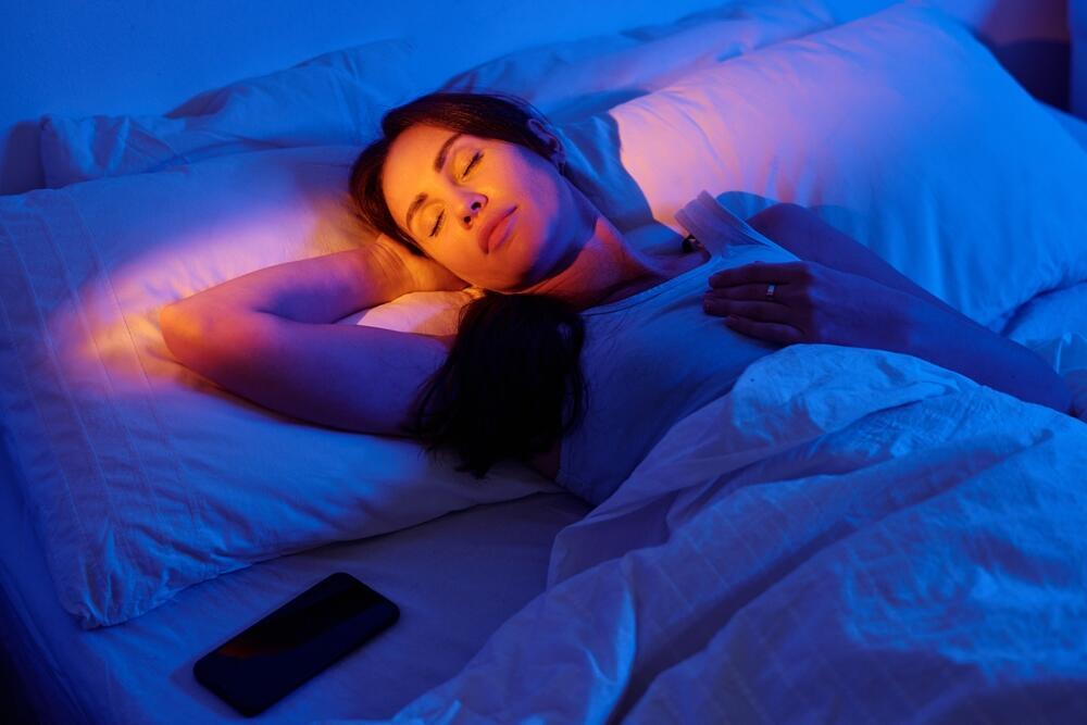
The harmful effects of even a tiny amount of light in the bedroom. Illustration
(Photo: shutterstock)
“Previous observational studies have linked nighttime light exposure with a higher risk of cardiovascular disease, but little is known about the related cardiac structure and functional changes,” Qi said. “We carried out this research to find out what happens to the heart over time when people are exposed to too much light at nighttime.”
The key innovation
To investigate, the researchers turned to the UK Biobank, a massive British database used for studies around the world. Although the study was led by U.S. researchers, the underlying data came from 11,071 British adults with an average age of 61, all of whom were free of cardiovascular disease at the outset. Each participant wore a sensor on the wrist that measured light and movement for seven consecutive days.
About three years later, the participants underwent cardiac magnetic resonance imaging, or MRI, considered the most accurate method for measuring the heart’s structure and function. It can detect subclinical changes before they develop into overt disease. This ability to effectively look inside the heart is what made the study novel. Until now, such imaging has primarily been used to examine the effects of other environmental hazards on the heart, such as air pollution and aircraft noise, rather than the effects of nighttime light.
Sleep
(Photo: shutterstock)
The threshold set by the researchers was 3 lux, a unit used to measure illuminance. That is very little light. It represents an almost completely dark room, not a brightly lit one. The choice was not arbitrary: It is roughly the intensity at which light begins to suppress secretion of melatonin, the hormone that regulates sleep.
“It’s still a very dim room. Your closed eyes, however, would be able to detect the light,” Kristen Knutson, an associate professor of sleep medicine at Northwestern University’s Feinberg School of Medicine.
Qi cited streetlight filtering through blinds, the glow of an alarm clock and indicator lights on household devices such as air purifiers and humidifiers as possible sources of such light. More than a quarter of participants had virtually no exposure to light of this intensity during sleeping hours, and they served as the comparison group.
What did the MRI scans reveal?
The MRI scans showed a consistent pattern among those with the highest exposure to nighttime light. The wall of the left ventricle, the heart’s main pumping chamber, was thicker and its muscle mass was greater, while the chamber itself was narrower. This combination is known as concentric hypertrophy, a recognized sign of a heart under chronic strain.
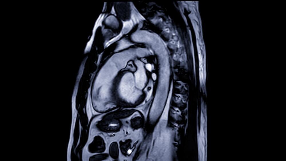
A view of the heart on an MRI scan
(Photo: shutterstock)
Compared with those who slept in near-total darkness, those exposed to the most nighttime light had an average 2.4% greater left ventricular muscle mass and 1.5% greater average wall thickness. At the same time, researchers found a 1.9% decline in the heart muscle’s ability to contract, along with declines in measures of how the muscle deforms with each heartbeat, early signs of impaired function before symptoms are felt. Related changes were also observed in the right ventricle and left atrium.
More importantly, the association followed a gradient: The greater the exposure to light, the more pronounced the changes in the heart.
“This is the first study of its kind, and it shows that exposure to higher levels of light at nighttime is linked to cardiac remodelling. This is where the structure and function of the heart changes and we typically see it in response to chronic stress or injury to the heart,” Qi said. “It’s our body’s way of adapting to that stress, but ultimately it weakens the heart and can lead to heart failure.”
Small in percentage, not in significance
At first glance, a change of 1% or 2% may sound negligible. The researchers sought to put those figures into perspective. The magnitude of the effect, they wrote, was similar to that found in the same database for well-established environmental risk factors, including exposure to fine particulate air pollution and aircraft noise. In other words, nighttime light leaves a mark on the heart comparable in magnitude to that associated with air pollution.
“Our findings, together with evidence from other studies, suggest that reduction of nighttime light exposure should be considered as one of the potential strategies for preventing heart disease by clinicians and policy makers,” Qi said.
That is where the broader context comes in. In a much larger group from the database, comprising more than 70,000 people followed for about eight to 10 years, those with the greatest exposure to nighttime light had a higher risk of heart failure, stroke, heart attack, atrial fibrillation and death from cardiovascular disease. That finding is not entirely new and is consistent with previous research showing a similar association. The contribution of the current study lies not in identifying the risk, but in providing a visible structural basis for it — visual evidence of changes in the heart that had previously only been suspected.
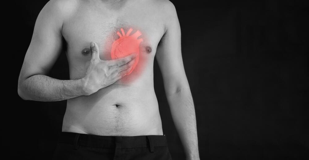
'Darkness deserves to be considered a vital sign, as important to heart health as controlling blood pressure and breathing clean air.'
(Photo: shutterstock)
The intuitive explanation is that light simply robs people of sleep and that insufficient sleep harms the heart. The researchers examined that possibility directly and found that it was only partly true. Their analysis showed that shorter sleep duration accounted for about 24% to 49% of the association between light exposure and changes in the heart, depending on the measure. In most cases, that means more than half of the effect appears to operate through entirely different pathways unrelated to sleep duration.
What are those pathways? This is where the body’s biological clock comes in. Light at night disrupts the circadian rhythm, the internal clock that synchronizes the body’s 24-hour cycles, from blood pressure and heart rate to hormone secretion and inflammatory processes. Such disruption has previously been linked to increased activity of the sympathetic nervous system, imbalances in stress hormones and impaired blood vessel function, all processes that can promote thickening of the heart muscle and impair its function.
“A future study about nighttime light could include people who start out with cardiovascular disease or are at high risk for it,” Knutson said.
Indirect evidence that the phenomenon is real rather than coincidental came from another part of the data. Exposure to bright light during daytime hours was barely associated with changes in the heart or cardiovascular events. That contrast makes biological sense because melatonin suppression is primarily triggered by light at night. It also reduces the likelihood that some external urban factor explains the findings.
Darkness may be as important as controlling blood pressure
An important caveat is needed. This was an observational study, meaning it shows an association but does not prove that light caused the changes in the heart. There is always the possibility of reverse causation: Perhaps people with less healthy hearts are more likely to sleep with the light on.
The researchers tried to address that possibility in several ways. First, they excluded anyone who already had heart disease at the outset. Second, when they repeated the analysis after excluding participants who underwent MRI scans less than two years after their light exposure was measured, the results remained consistent. Third, among a group of 951 participants who had undergone MRI scans before their light exposure was measured, their initial heart health did not predict how much light they were subsequently exposed to. Together, those three analyses weaken the reverse-causation explanation, though they do not eliminate it.
The study had other limitations. Light exposure was measured over just one week, researchers measured light intensity rather than its wavelength composition and the vast majority of participants were white, so it is unclear whether the findings apply to more diverse populations. Night-shift workers were excluded from the analysis for methodological reasons, meaning the conclusions are particularly limited when it comes to that group.

Next time, make sure your bedroom is completely dark. Illustration
(Photo: shutterstock)
An accompanying editorial was also published in the European Heart Journal, in which Prof. Thomas Münzel, an environmental cardiologist at University Medical Center Mainz in Germany, translated the findings into practical recommendations. He said it was time for doctors, particularly those treating patients with heart failure and atrial fibrillation, to ask their patients how dark their bedrooms are, whether they work night shifts and how much exposure they have to screens after dark.
His recommendations were simple and inexpensive: blackout curtains, warm-colored night lights and covering the small LED indicator lights on electronic devices.
Münzel went beyond the individual bedroom, arguing that light pollution is one of the few environmental hazards that cities can control directly and at relatively low cost.
“Ultimately, this study is more than an interesting observation,” he wrote. “It provides a crucial link between what satellites already show us, i.e. a planet glowing ever brighter at night, and the clinical reality unfolding inside our chests: hearts that are becoming stiffer, weaker, and less efficient. The relationship is biologically coherent, the effect sizes are clinically meaningful, and the dose–response is unambiguous: more light, worse outcomes. The takeaway is clear, darkness deserves recognition as a vital sign, as essential to cardiovascular health as blood pressure control and clean air.”